LIFE PROCESSES
WHAT ARE LIFE PROCESSES?
LIFE PROCESSES
NUTRITION
It is needed for body growth, development and synthesis of protein and other substances.
Even we are inactive, energy is needed to maintain a state of order in the body.
General requirement for energy & materials is common in all organisms, but it is fulfilled in different ways.
Some organisms use simple food material obtained from inorganic sources such as CO2 and water. They are called autotrophs. E.g. green plants and some bacteria.
Some organisms directly or indirectly depend on autotrophs for nutrition. They are called heterotrophs. E.g. animals and fungi. They utilise complex substances which are broken down into simpler ones with the help of biocatalysts called enzymes.
NUTRITION
Autotrophic Nutrition
It is a process where an organism prepares its own food from simple inorganic materials.
Photosynthesis: It is the process by which CO2 & water is converted into carbohydrates in presence of sunlight & chlorophyll. It occurs in autotrophs like green plants.


Carbohydrates provide energy to the plant.
The carbohydrates which are not used immediately are stored as starch. It serves as the internal energy reserve.
In our body, some of the energy derived from food is stored as glycogen.
A cross-section of a leaf under the microscope shows that some cells contain green dots. They are cell organelles called chloroplasts. It contains chlorophyll.
Events in photosynthesis:
- Absorption of light energy by chlorophyll.
- Conversion of light energy to chemical energy and splitting of water molecules into hydrogen & oxygen.
- Reduction of CO2 to carbohydrates.
These steps need not occur in sequence immediately. E.g. desert plants take up CO2 at night and prepare an intermediate. It is acted upon by the energy absorbed by chlorophyll during the day.
Experiment to show that chlorophyll is essential for photosynthesis:

- Take a potted plant with variegated leaves (e.g. money plant or crotons).
- Keep it in a dark room for 3 days to use up all the starch (destarch).
- Keep the plant in sunlight for about 6 hours.
- Pluck a leaf and mark the green areas in it and trace them on a paper.
- Dip the leaf in boiling water for a few minutes.
- Then immerse it in a beaker containing alcohol. Place the beaker in a water-bath and heat to boil the alcohol.
- The leaf becomes colourless. Chlorophyll is dissolved in alcohol and the alcohol turns green.
- Dip the leaf in a dilute iodine solution for a few minutes.
- The green areas of leaf turn dark blue. It indicates the presence of starch. Colourless part of leaf shows no formation of starch.
Gaseous exchange occurs in the leaves through stomata for photosynthesis. It also occurs across the surface of stems, roots and leaves.
During this, much water is also lost through stomata. So the plant closes stomata when it does not need CO2.
The opening and closing of stomatal pore is regulated by guard cells. When water flows into guard cells, they swell and the pore opens. If the guard cells shrink, the pore closes.

Experiment to show that CO2 is essential for photosynthesis:

- Take 2 healthy potted plants having nearly same size.
- Keep them in a dark room for 3 days.
- Now place each plant on separate glass plates. In one, place a watch-glass containing potassium hydroxide (KOH). KOH is used to absorb CO2.
- Cover both plants with separate bell-jars.
- Using Vaseline, seal the bottom of the jars to the glass plates to make it air-tight.
- Keep the plants in sunlight for about two hours.
- Test the leaves from both plants using iodine.
- Leaf of plant kept without KOH turn blue. It indicates the presence of starch. Plant with KOH does not turn blue.
Experiment to show that sunlight is essential for photosynthesis:
- Keep a plant in dark room for 3 days to destarch leaves.
- Cover a leaf partially with a black paper.
- Expose the plant to sunlight for 3-4 hours.
- Remove chlorophyll from the leaf and perform a starch test with iodine.
- Covered leaf part shows brown colour. Exposed leaf turns dark blue due to the presence of starch.
Autotrophs also need other raw materials such as water and minerals like N, P, Fe and Mg. They are taken up from the soil. Nitrogen is an essential element for protein synthesis. It is absorbed as inorganic nitrates or nitrites. Or it is taken up as organic compounds prepared by bacteria from atmospheric nitrogen.Heterotrophic Nutrition
It is the nutrition in which an organism depends on other living organisms for food.
Organisms take and use food in various ways. E.g.- Some organisms break down food outside the body and absorb it. E.g. fungi like bread moulds, yeast and mushrooms.
- Some take in food and break down inside the body.
- Some organisms take food from organisms without killing them (parasitism). E.g., Cuscuta (amar-bel), ticks, lice, leeches and tape-worms.

Events in photosynthesis:
- Absorption of light energy by chlorophyll.
- Conversion of light energy to chemical energy and splitting of water molecules into hydrogen & oxygen.
- Reduction of CO2 to carbohydrates.
Experiment to show that chlorophyll is essential for photosynthesis:
- Take a potted plant with variegated leaves (e.g. money plant or crotons).
- Keep it in a dark room for 3 days to use up all the starch (destarch).
- Keep the plant in sunlight for about 6 hours.
- Pluck a leaf and mark the green areas in it and trace them on a paper.
- Dip the leaf in boiling water for a few minutes.
- Then immerse it in a beaker containing alcohol. Place the beaker in a water-bath and heat to boil the alcohol.
- The leaf becomes colourless. Chlorophyll is dissolved in alcohol and the alcohol turns green.
- Dip the leaf in a dilute iodine solution for a few minutes.
- The green areas of leaf turn dark blue. It indicates the presence of starch. Colourless part of leaf shows no formation of starch.

Experiment to show that CO2 is essential for photosynthesis:

- Take 2 healthy potted plants having nearly same size.
- Keep them in a dark room for 3 days.
- Now place each plant on separate glass plates. In one, place a watch-glass containing potassium hydroxide (KOH). KOH is used to absorb CO2.
- Cover both plants with separate bell-jars.
- Using Vaseline, seal the bottom of the jars to the glass plates to make it air-tight.
- Keep the plants in sunlight for about two hours.
- Test the leaves from both plants using iodine.
- Leaf of plant kept without KOH turn blue. It indicates the presence of starch. Plant with KOH does not turn blue.
Experiment to show that sunlight is essential for photosynthesis:
- Keep a plant in dark room for 3 days to destarch leaves.
- Cover a leaf partially with a black paper.
- Expose the plant to sunlight for 3-4 hours.
- Remove chlorophyll from the leaf and perform a starch test with iodine.
- Covered leaf part shows brown colour. Exposed leaf turns dark blue due to the presence of starch.
Heterotrophic Nutrition
- Some organisms break down food outside the body and absorb it. E.g. fungi like bread moulds, yeast and mushrooms.
- Some take in food and break down inside the body.
- Some organisms take food from organisms without killing them (parasitism). E.g., Cuscuta (amar-bel), ticks, lice, leeches and tape-worms.
HOW DO ORGANISMS OBTAIN THEIR NUTRITION?
Unicellular organisms take food by their entire surface.
As the complexity increases, specialised parts are formed to perform different functions.
E.g., Amoeba captures food using temporary finger-like extensions (Pseudopodia) of the cell surface. They fuse over the food particle forming a food-vacuole. In this, food breaks down into simpler ones and diffuse into cytoplasm. The undigested material is moved to cell surface and thrown out.

Paramoecium takes food in at a specific spot by the movement of cilia (hair-like structure covering the cell).
Nutrition in Human Beings
Human alimentary canal is a long tube extending from the mouth to the anus.

Food digestion:
It is the breakdown of complex food with the help of enzymes into smaller absorbable molecules.
In mouth, food is chewed with the help of teeth and muscular tongue and it is mixed with saliva secreted by the salivary glands.
Teeth are used to crush the food into small particles.
Saliva contains salivary amylase enzyme that breaks down starch to simple sugar.
Experiment to show the action of saliva:- Take 1 mL starch solution (1%) in test tubes A & B.
- Add 1 mL saliva to test tube A and leave both test tubes undisturbed for 20-30 minutes. Now add a few drops of dilute iodine solution to the test tubes.
- In test tube B, blue colour develops. It indicates presence of starch. In test-tube A, no colour change occurs because starch is digested by salivary amylase.
The lining of the alimentary canal is soft for smooth passage of food. The muscles in the lining, contract rhythmically to push the food forward. It is called peristalsis. It helps to process the food properly in each part.
From the mouth, food is moved through oesophagus (food-pipe) and reaches stomach.
Stomach is a large organ that expands when food enters it. Its muscular walls help in mixing the food thoroughly with digestive juices.
Stomach wall contains gastric glands. They release hydrochloric acid, pepsin (a protein digesting enzyme) and mucus.
HCl creates an acidic medium which facilitates the action of pepsin. It also helps to destroy harmful bacteria.
The mucus protects the inner lining of the stomach from the action of the acid.
Sometimes HCl acts on the wall causing a burning sensation. This is called acidity.
A sphincter muscle regulates the exit of food in small amounts from stomach into small intestine.
Small intestine is the longest and highly coiled part of the alimentary canal.
In animals, the length of the small intestine differs based on the food they eat. E.g.- Herbivores have a longer small intestine for cellulose digestion.
- Meat is easier to digest. So carnivores have a shorter small intestine.
Small intestine is the site of the complete digestion of carbohydrates, proteins & fats. It receives the secretions of the liver & pancreas.
Liver secretes bile juice. It- Emulsifies fat. It is the breakdown of large fat globules into the smaller globules with the help of bile salts. It increases the efficiency of enzyme action. This is similar to emulsifying action of soaps on dirt.
- Makes the acidic food (from stomach) alkaline.
Pancreas secretes pancreatic juice. It contains enzymes like trypsin (to digest proteins) and lipase (to digest emulsified fats).
Glands in the walls of small intestine secrete intestinal juice which contains enzymes. They finally convert- Proteins → amino acids.
- Complex carbohydrates → glucose.
- Fats → fatty acids & glycerol.
Digested food is taken up by the walls of the intestine.
Inner lining of the small intestine has many finger-like projections called villi. They increase the surface area for absorption. Villi contain blood vessels that take the absorbed food to all cells. In cells, it is utilised to obtain energy, build up new tissues and repair old tissues.
The unabsorbed food is moved to the large intestine where its wall absorbs more water from this material.
Remaining waste material is removed from the body via anus. This removal is regulated by anal sphincter.Dental caries (Tooth decay):
It is the gradual softening & demineralisation of enamel & dentine due to the production of acids by bacteria. They convert sugary foods to acids.
Bacteria invade the pulp causing inflammation & infection.
Masses of bacterial cells together with food particles stick to the teeth to form dental plaque. It prevents saliva reaching the tooth surface to neutralise the acid.
Brushing the teeth after eating can remove plaque.

Nutrition in Human Beings
Human alimentary canal is a long tube extending from the mouth to the anus.

Food digestion:
- Take 1 mL starch solution (1%) in test tubes A & B.
- Add 1 mL saliva to test tube A and leave both test tubes undisturbed for 20-30 minutes. Now add a few drops of dilute iodine solution to the test tubes.
- In test tube B, blue colour develops. It indicates presence of starch. In test-tube A, no colour change occurs because starch is digested by salivary amylase.
- Herbivores have a longer small intestine for cellulose digestion.
- Meat is easier to digest. So carnivores have a shorter small intestine.
- Emulsifies fat. It is the breakdown of large fat globules into the smaller globules with the help of bile salts. It increases the efficiency of enzyme action. This is similar to emulsifying action of soaps on dirt.
- Makes the acidic food (from stomach) alkaline.
- Proteins → amino acids.
- Complex carbohydrates → glucose.
- Fats → fatty acids & glycerol.
Dental caries (Tooth decay):
It is the gradual softening & demineralisation of enamel & dentine due to the production of acids by bacteria. They convert sugary foods to acids.
Bacteria invade the pulp causing inflammation & infection.
Masses of bacterial cells together with food particles stick to the teeth to form dental plaque. It prevents saliva reaching the tooth surface to neutralise the acid.
Brushing the teeth after eating can remove plaque.
LIFE PROCESSES
RESPIRATION
Some organisms use oxygen to breakdown glucose into CO2 and water. Some do not use oxygen.
Experiment to prove release of CO2 during respiration in human
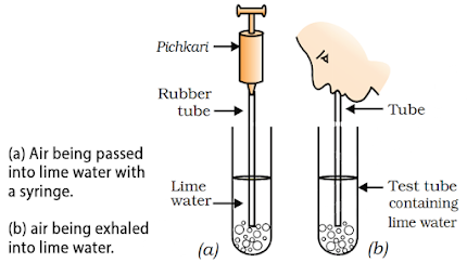
- Take some freshly prepared lime water in 2 test tubes.
- In one, blow air through lime water. It immediately turns lime water milky.
- In other test tube, pass air using a syringe or pichkari. It takes much time to turn the lime water milky.
- It shows that the breath-out air contains more CO2 as compared to atmospheric air.
Experiment to prove release of CO2 during respiration in Yeast
- Add some yeast to fruit juice or sugar solution. Take this mixture in a test tube fitted with a one-holed cork.
- Fit the cork with a bent glass tube. Dip its free end into a test tube containing freshly prepared lime water.
- Air taken out through the tube makes lime water milky. It is due to the production of CO2 in the mixture of yeast & sugar solution. Here, fermentation occurs.
In all types of respiration, the first step is the breakdown of glucose (6-carbon) into pyruvate (3-carbon). It takes place in the cytoplasm.

Anaerobic respiration: It is the respiration in the absence of air (oxygen). It releases less energy. E.g.- In yeast, the pyruvate is converted into ethanol & CO2. It occurs during fermentation.
- Sometimes, when there is no oxygen in our muscle cells, the pyruvate breaks down into lactic acid (3-carbon). This build-up of lactic acid in muscles during sudden activity causes cramps.
Aerobic respiration: It is the respiration in presence of air (oxygen). It releases much energy. Here, pyruvate breaks down using oxygen in the mitochondria giving three CO2 molecules & water.Energy released during cellular respiration is used immediately to synthesise ATP molecules (energy currency) from ADP & inorganic phosphate [℗].

ATP is used to fuel all other cellular activities. When the terminal phosphate linkage in ATP is broken using water, energy (30.5 kJ/mol) is released. It drives the endothermic reactions in the cell.
A battery is used to obtain mechanical energy, light energy, electrical energy etc. Similarly, ATP can be used for muscle contraction, protein synthesis, conduction of nervous impulses etc.
Gas exchange in plants:
It occurs through stomata. Here, CO2 & oxygen are exchanged by diffusion. The large intercellular spaces in leaves help the cells in contact with air.
During day, CO2 formed by respiration is used for photosynthesis. So, CO2 is not released but oxygen is released. At night, photosynthesis does not occur. So, CO2 is released out but oxygen is not released.
Gas exchange in animals:
Aquatic animals breathe dissolved oxygen in water.
In fishes, the respiratory organ is gills with gill slits behind their eyes. They may be covered by operculum.
During breathing, fishes open and close mouth & gill slits (or operculum) in a coordinated manner and timing. They take in water through mouth and pumps over the gills. From the gills, dissolved O2 is taken up by blood.
The amount of dissolved O2 is lower than that in the air. So, the rate of breathing in aquatic organisms is faster than that in terrestrial organisms.
In terrestrial animals, there are different types of organs to breathe atmospheric oxygen. They increase surface area which is in contact with the atmosphere.
Surface of respiratory organs is very fine and delicate for easy gas exchange. To protect this surface, it is placed within the body. So, some passages are necessary to carry air in and out of respiratory organ.
Human respiratory system
It involves lungs & air passage.
Air passage starts from nostrils through which air is taken into the body. Air passage is lined with fine hairs & mucus to filter the air.
Conduction of air through the passage is as follows:
Nostrils → nasal passage → pharynx → larynx → trachea → bronchi → bronchioles → lungs.
Rings of cartilage in the throat and trachea prevent collapsing of air-passage.

Within the lungs, the passage divides into smaller tubes (bronchi & bronchioles).
Bronchioles terminate in balloon-like structures called alveoli (sing. alveolus).
Gas exchange occurs in the surface alveoli.
Alveolar walls contain a network of blood vessels.
When we breathe in, ribs are lifted and the diaphragm gets flattened. As a result, the chest cavity becomes larger and air enters the lung alveoli.
The blood brings CO2 from the rest of the body to release into the alveoli. Oxygen in the alveoli is taken up by blood in the alveolar blood vessels and is transported to all body parts.Tobacco or tobacco products affect tongue, lungs, heart and liver. Smokeless tobacco also causes heart attacks, strokes, pulmonary diseases & cancers.
Oral cancer is highly reported in India due to tobacco chewing in the form of gutkha.
Smoking destroys cilia on the upper respiratory tract. As a result, germs, dust, smoke etc. enter lungs and cause infection, cough & lung cancer (common cause of death).
During the breathing cycle, the lungs always contain a residual volume of air so that there is sufficient time to absorb oxygen and release CO2.
In large-sized animals, diffusion pressure is not sufficient to deliver O2 to all body parts. So, respiratory pigments take up oxygen from the lungs and carry it to tissues.
In humans, the respiratory pigment is haemoglobin on red blood corpuscles (RBC). It has high affinity for O2.
CO2 is more soluble in water than oxygen and hence is mostly transported in the dissolved form in our blood.
- If the alveolar surface were spread out, it would cover about 80 m2. The surface area of human body is about 1.9 m2.
- If diffusion were to move oxygen in our body, it would take 3 years for an oxygen molecule to get to toes from our lungs. Haemoglobin helps in faster gas transport.
RESPIRATION
Experiment to prove release of CO2 during respiration in human

- Take some freshly prepared lime water in 2 test tubes.
- In one, blow air through lime water. It immediately turns lime water milky.
- In other test tube, pass air using a syringe or pichkari. It takes much time to turn the lime water milky.
- It shows that the breath-out air contains more CO2 as compared to atmospheric air.
Experiment to prove release of CO2 during respiration in Yeast
- Add some yeast to fruit juice or sugar solution. Take this mixture in a test tube fitted with a one-holed cork.
- Fit the cork with a bent glass tube. Dip its free end into a test tube containing freshly prepared lime water.
- Air taken out through the tube makes lime water milky. It is due to the production of CO2 in the mixture of yeast & sugar solution. Here, fermentation occurs.

- In yeast, the pyruvate is converted into ethanol & CO2. It occurs during fermentation.
- Sometimes, when there is no oxygen in our muscle cells, the pyruvate breaks down into lactic acid (3-carbon). This build-up of lactic acid in muscles during sudden activity causes cramps.

Gas exchange in plants:
It occurs through stomata. Here, CO2 & oxygen are exchanged by diffusion. The large intercellular spaces in leaves help the cells in contact with air.
During day, CO2 formed by respiration is used for photosynthesis. So, CO2 is not released but oxygen is released. At night, photosynthesis does not occur. So, CO2 is released out but oxygen is not released.
Gas exchange in animals:
Aquatic animals breathe dissolved oxygen in water.
In fishes, the respiratory organ is gills with gill slits behind their eyes. They may be covered by operculum.
During breathing, fishes open and close mouth & gill slits (or operculum) in a coordinated manner and timing. They take in water through mouth and pumps over the gills. From the gills, dissolved O2 is taken up by blood.
The amount of dissolved O2 is lower than that in the air. So, the rate of breathing in aquatic organisms is faster than that in terrestrial organisms.
In terrestrial animals, there are different types of organs to breathe atmospheric oxygen. They increase surface area which is in contact with the atmosphere.
Surface of respiratory organs is very fine and delicate for easy gas exchange. To protect this surface, it is placed within the body. So, some passages are necessary to carry air in and out of respiratory organ.
Human respiratory system

Tobacco or tobacco products affect tongue, lungs, heart and liver. Smokeless tobacco also causes heart attacks, strokes, pulmonary diseases & cancers.During the breathing cycle, the lungs always contain a residual volume of air so that there is sufficient time to absorb oxygen and release CO2.
Oral cancer is highly reported in India due to tobacco chewing in the form of gutkha.
Smoking destroys cilia on the upper respiratory tract. As a result, germs, dust, smoke etc. enter lungs and cause infection, cough & lung cancer (common cause of death).
- If the alveolar surface were spread out, it would cover about 80 m2. The surface area of human body is about 1.9 m2.
- If diffusion were to move oxygen in our body, it would take 3 years for an oxygen molecule to get to toes from our lungs. Haemoglobin helps in faster gas transport.
LIFE PROCESSES
TRANSPORTATION
It is the process of movement of water and other molecules to the concerned parts of the organism.
Transportation in Human Beings
Human circulatory system: Blood, Heart & Blood vessels.
Blood
Blood is a fluid connective tissue.
It consists of a fluid medium called plasma in which blood cells (RBC, WBC & platelets) are suspended.
Plasma transports food, O2, CO2 and nitrogenous wastes.
Oxygen is mainly transported by Haemoglobin.
Normal level of haemoglobin in human beings:- In men: 14 to 17 g/ 100 ml.
- In women: 12 to 15 g/ 100 ml.
- In children: 11 to 16 g/ 100 ml.
The normal level of haemoglobin in animals like buffalo or cow is 10.4 to 16.4 g/ 100 ml. Haemoglobin content in calves is higher than male and female animals.
Adult men do more work than women and children. So they need more oxygen to get energy. That’s why adult men have more haemoglobin.
Haemoglobin level in human is comparatively more than that of animals like cattle because human body needs more oxygen to do various biological works.
Our pump – the heart A schematic sectional view of the human heartHeart pumps the blood all over the body.
It is a muscular organ that is as big as our fist.
It has 4 chambers: 2 upper right and left atria and 2 lower right and left ventricles.
Right chambers carry CO2-rich (deoxygenated) blood. Left chambers carry O2-rich (oxygenated) blood.
Deoxygenated blood reaches the lungs to remove CO2. Oxygenated blood from the lungs is brought back to the heart and then pumped to the rest of the body.
Pumping process of heart:
- Oxygenated blood from the lungs → left atrium relaxes → blood enters left atrium → left atrium contracts & left ventricle relaxes → blood enters left ventricle → left ventricle contracts → blood is pumped out to the body.
- Deoxygenated blood from the body → right atrium relaxes → blood enters right atrium → right atrium contracts & right ventricle dilates → blood transfers to right ventricle → right ventricle contracts → blood is pumped into the lungs for oxygenation.
Since ventricles have to pump blood into various organs, they have thicker muscular walls than that of atria.
Heart has valves to prevent the backflow of blood when the atria or ventricles contract.

Schematic representation of transport and exchange of oxygen and CO2
Oxygen enters the blood in the lungs
The separation of right side and left side of the heart prevents mixing of oxygenated and deoxygenated blood. This allows a highly efficient supply of oxygen to the body. It is useful in animals that need high energy (birds & mammals) to maintain body temperature.
Animals like amphibians & many reptiles do not use energy to maintain temperature. They depend on the temperature in the environment. Such animals have 3-chambered heart, and tolerate some mixing of the oxygenated and deoxygenated blood.
Fishes have only 2-chambered heart. Here, circulation occurs as follows:
Deoxygenated blood enters the heart → pumped to gills → blood is oxygenated in gills → blood to rest of the body.
Thus, blood goes only once through the heart during one cycle of passage through the body.
In other vertebrates, blood goes through the heart twice during each cycle. This is called double circulation.
Blood pressure (BP)
- It is the force that blood exerts against the wall of a vessel. This is much greater in arteries than in veins.
- Systolic pressure: Blood pressure in the artery during ventricular systole (contraction). It is about 120 mm Hg.
- Diastolic pressure: Blood pressure in the artery during ventricular diastole (relaxation). It is about 80 mm Hg.
- Sphygmomanometer: An instrument to measure BP.
- High BP (hypertension) is caused by the constriction of arterioles, which increases resistance to blood flow. It leads to the rupture of an artery and internal bleeding.
TRANSPORTATION
It is the process of movement of water and other molecules to the concerned parts of the organism.
Transportation in Human Beings
Human circulatory system: Blood, Heart & Blood vessels.
Blood
- In men: 14 to 17 g/ 100 ml.
- In women: 12 to 15 g/ 100 ml.
- In children: 11 to 16 g/ 100 ml.

Pumping process of heart:
- Oxygenated blood from the lungs → left atrium relaxes → blood enters left atrium → left atrium contracts & left ventricle relaxes → blood enters left ventricle → left ventricle contracts → blood is pumped out to the body.
- Deoxygenated blood from the body → right atrium relaxes → blood enters right atrium → right atrium contracts & right ventricle dilates → blood transfers to right ventricle → right ventricle contracts → blood is pumped into the lungs for oxygenation.
Since ventricles have to pump blood into various organs, they have thicker muscular walls than that of atria.
Heart has valves to prevent the backflow of blood when the atria or ventricles contract.

Schematic representation of transport and exchange of oxygen and CO2
Oxygen enters the blood in the lungs
The separation of right side and left side of the heart prevents mixing of oxygenated and deoxygenated blood. This allows a highly efficient supply of oxygen to the body. It is useful in animals that need high energy (birds & mammals) to maintain body temperature.
Animals like amphibians & many reptiles do not use energy to maintain temperature. They depend on the temperature in the environment. Such animals have 3-chambered heart, and tolerate some mixing of the oxygenated and deoxygenated blood.
Fishes have only 2-chambered heart. Here, circulation occurs as follows:
Deoxygenated blood enters the heart → pumped to gills → blood is oxygenated in gills → blood to rest of the body.
Thus, blood goes only once through the heart during one cycle of passage through the body.
In other vertebrates, blood goes through the heart twice during each cycle. This is called double circulation.
Blood pressure (BP)
- It is the force that blood exerts against the wall of a vessel. This is much greater in arteries than in veins.
- Systolic pressure: Blood pressure in the artery during ventricular systole (contraction). It is about 120 mm Hg.
- Diastolic pressure: Blood pressure in the artery during ventricular diastole (relaxation). It is about 80 mm Hg.
- Sphygmomanometer: An instrument to measure BP.
- High BP (hypertension) is caused by the constriction of arterioles, which increases resistance to blood flow. It leads to the rupture of an artery and internal bleeding.
The tubes – blood vessels
It includes arteries, veins and capillaries.
Arteries: They carry blood from heart to various body parts. Since the blood emerges from the heart under high pressure, the arteries have thick, elastic walls.On reaching an organ or tissue, the artery divides into small branches (arterioles) to bring the blood in contact with all the cells.
Veins: They collect the blood from different organs and bring it back to the heart. They have no thick walls because the blood is no longer under pressure. Instead, they have valves to flow the blood only in one direction.
Capillaries: The smallest vessels having walls which are one-cell thick. Through this wall, exchange of material between blood and surrounding cells takes place.The capillaries join together to form veins that convey the blood away from the organ or tissue.
Maintenance by platelets
Leakage or loss of blood due to injury leads to reduction in pressure and efficiency of circulatory system.
To avoid this, the platelet cells plug these leaks to clot the blood at the points of injury.
Lymph (Tissue fluid):
Through the pores in the capillary walls, some amount of plasma, proteins and blood cells escape into intercellular spaces in the tissues to form lymph.
It is similar to blood plasma but colourless and contains less protein.
From intercellular spaces, lymph drains into lymphatic capillaries, which join to large lymph vessels that finally open into larger veins.
Lymph carries digested fat from intestine and drains excess fluid from extracellular space back into the blood.
Transportation in Plants
Soil is the nearest and richest source of raw materials (water & minerals).
If the distance between roots & leaves are small, energy and raw materials can easily diffuse to all parts. But if the distance is large, a transportation system is essential.
Plants do not move and have enormous dead cells. So, they need only low energy and slow transport systems.
Plant transport systems include 2 independently organised conducting tubes:- Xylem: It moves water & minerals from the soil.
- Phloem: It transports products of photosynthesis (energy stores) from leaves to other parts.
Transport of water
In xylem tissue, vessels & tracheids of the roots, stems and leaves are interconnected to form water-conducting channels reaching all parts.
At the roots, cells actively take up ions from soil. This creates a difference in the concentration of ions between root and soil. So, water moves into the root from the soil. Thus there is steady movement of water into root xylem, creating a column of water pushing upwards.
But this pressure is not enough to move water over the heights. So, plants use another strategy called transpiration.
Transpiration is the loss of water vapour from the aerial parts (mainly stomata of leaves) of the plant. It can be proved by the following activity.- Take two small same sized pots with same amount of soil. One should have a plant in it. In other pot, place a stick of the same height as the plant.
- Cover the soil in both pots with a plastic sheet so that moisture cannot escape by evaporation.
- Cover both sets with plastic sheets and place in bright sunlight for half an hour.
- In pot with plant, water droplets are found in plastic sheet. It is due to condensation of water vapour released by transpiration. In other pot, water droplets are not formed.
The water which is lost through the stomata is replaced by water from the xylem vessels in the leaf.
Transpiration creates a suction which pulls water from the xylem cells of roots.
Thus, transpiration helps in the absorption and upward movement of water & minerals from roots to the leaves. It also helps in temperature regulation.
Generally, stomata are closed at night. So, plants depend on the root pressure for water transport at night.
During the day when stomata are open, the transpiration pull is the major driving force in the water transport.Transport of food & other substances
Transport of soluble products of photosynthesis (food) from leaves to other parts is called translocation.
It occurs in a vascular tissue called phloem.
Phloem also transports amino acids & other substances.
These substances are delivered to the storage organs of roots, fruits and seeds and to growing organs.
Translocation takes place in the sieve tubes with the help of adjacent companion cells both in upward and downward directions.
Xylem transport occurs mainly by simple physical forces. But for the translocation in phloem, energy is utilised.
Material like sucrose is transferred into phloem using energy from ATP. This increases the osmotic pressure of the tissue causing water to move into it. This pressure moves the material in the phloem to tissues. E.g., in the spring, sugar stored in root or stem is transported to the buds which need energy to grow.
Maintenance by platelets
Leakage or loss of blood due to injury leads to reduction in pressure and efficiency of circulatory system.
To avoid this, the platelet cells plug these leaks to clot the blood at the points of injury.
Lymph (Tissue fluid):
Through the pores in the capillary walls, some amount of plasma, proteins and blood cells escape into intercellular spaces in the tissues to form lymph.
It is similar to blood plasma but colourless and contains less protein.
From intercellular spaces, lymph drains into lymphatic capillaries, which join to large lymph vessels that finally open into larger veins.
Lymph carries digested fat from intestine and drains excess fluid from extracellular space back into the blood.
Transportation in Plants
- Xylem: It moves water & minerals from the soil.
- Phloem: It transports products of photosynthesis (energy stores) from leaves to other parts.
Transport of water
- Take two small same sized pots with same amount of soil. One should have a plant in it. In other pot, place a stick of the same height as the plant.
- Cover the soil in both pots with a plastic sheet so that moisture cannot escape by evaporation.
- Cover both sets with plastic sheets and place in bright sunlight for half an hour.
- In pot with plant, water droplets are found in plastic sheet. It is due to condensation of water vapour released by transpiration. In other pot, water droplets are not formed.
Transport of food & other substances
LIFE PROCESSES
EXCRETION
EXCRETION
Excretion is the process of removal of harmful metabolic nitrogenous wastes from the body.
Excretion in Human Beings
Human excretory system includes a pair of kidneys, a pair of ureters, a urinary bladder & a urethra.

Kidneys are located in the abdomen, one on either side of the backbone.
The purpose of making urine is to filter out waste products from the blood.
In the kidneys, nitrogenous wastes such as urea or uric acid are removed from blood.
Each kidney has large numbers of filtration units called nephrons packed close together.

At the end of each nephron, a cup-shaped structure called Bowman’s capsule is seen. It encloses a cluster of very thin-walled blood capillaries called glomerulus.
In glomerulus, blood is filtered and the Bowman’s capsule collects the filtrate.
Glucose, amino acids, salts & major amount of water in the initial filtrate are selectively reabsorbed as the urine flows along the tube.
In a healthy adult, the initial filtrate is about 180 L daily. However, only 1-2 litre/day is excreted out because the remaining filtrate is reabsorbed in the kidney tubules.
Water is reabsorbed based on amount of excess water in the body and amount of dissolved waste is to be excreted.
The urine from each kidney enters a long tube (ureter), which then passes to urinary bladder and stored in it.
As the bladder expands, pressure increases that leads to the urge to urinate through the urethra.
The bladder is muscular, so it is under nervous control. As a result, we can control the urge to urinate.
Artificial kidney (Hemodialysis)
- Kidney failure leads to accumulation of poisonous wastes in the body. In this case, an artificial kidney can be used. It is a device to remove nitrogenous wastes from the blood through dialysis.
- Artificial kidneys contain many semi-permeable tubes suspended in a tank filled with dialysing fluid (it has same osmotic pressure of blood, but no nitrogenous wastes).
- When patient’s blood is passed through these tubes, the waste products from blood diffuses into dialysing fluid.

- The purified blood is pumped back into the patient.
- This is similar to the function of the kidney, but there is no reabsorption involved.
Organ donation
Any people can donate their organ or tissue regardless of age or gender.
Transplantation is required when recipient’s organ has been damaged or failed by disease or injury.
In organ transplantation, the organ is surgically removed from one person (organ donor) and transplanted to another person (recipient).
Most organ and tissue donations occur just after the death or brain death of the donor. But some organs (kidney, part of a liver, lung, etc.) and tissues can be donated while the donor is alive.